Understanding Pelger-Huët Cells: Causes and Implications

Pelger-Huët cells are a rare and fascinating phenomenon in hematology, characterized by abnormal neutrophil morphology. These cells, often discovered incidentally during routine blood smear examinations, can raise concerns due to their resemblance to immature or dysplastic cells. Understanding the causes and implications of Pelger-Huët cells is crucial for both healthcare professionals and patients. This condition, though typically benign, can sometimes mimic more serious disorders, making accurate diagnosis essential. By exploring the underlying genetics, clinical significance, and diagnostic approaches, this post aims to provide clarity on Pelger-Huët cells and their role in medical assessments. (Pelger-Huët anomaly, neutrophil morphology, hematology)
What Are Pelger-Huët Cells?

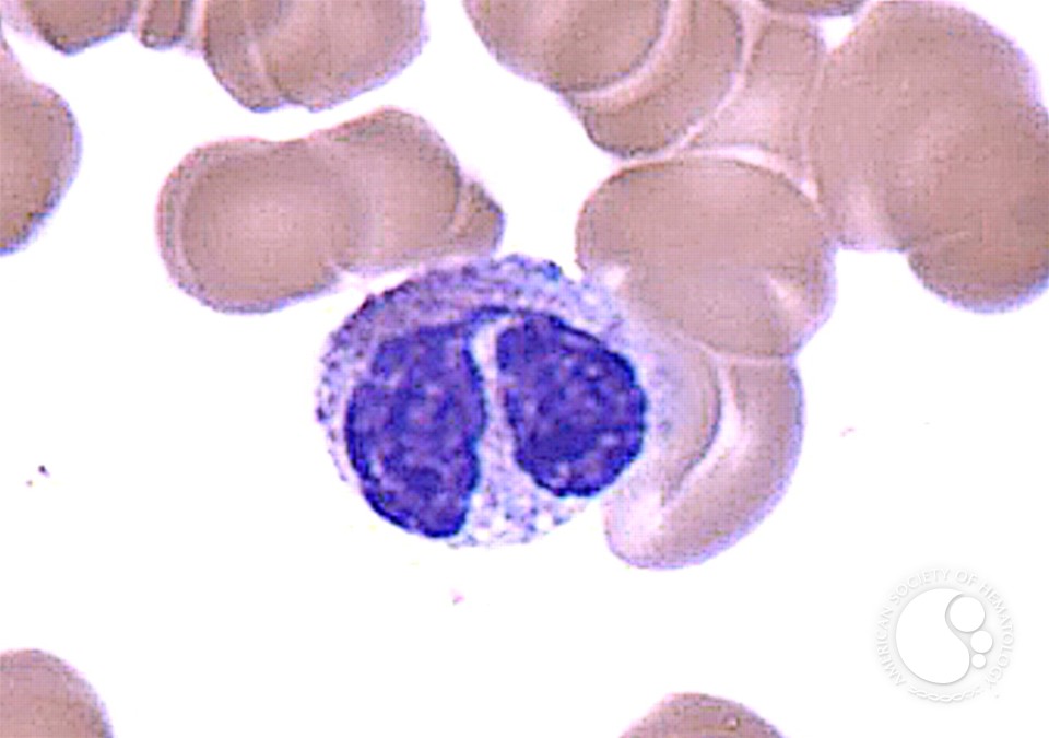
Pelger-Huët cells are neutrophils with abnormal nuclear segmentation, appearing as bilobed or bilobular nuclei instead of the typical multilobed structure. This condition, known as the Pelger-Huët anomaly, is usually hereditary and caused by mutations in the LBR gene. While these cells are often asymptomatic, their presence can complicate blood smear interpretations, leading to potential misdiagnosis. Recognizing Pelger-Huët cells is vital to differentiate them from conditions like myelodysplastic syndrome or leukemia. (Pelger-Huët anomaly, neutrophil morphology, hematology)
Causes of Pelger-Huët Cells

Genetic Basis
The primary cause of Pelger-Huët cells is a mutation in the LBR gene, which encodes a protein involved in nuclear envelope formation. This mutation disrupts neutrophil maturation, resulting in abnormal nuclear morphology. The condition is typically inherited in an autosomal dominant manner, meaning one copy of the mutated gene is sufficient to cause the anomaly. (genetic mutation, LBR gene, autosomal dominant)
Acquired Forms
While rare, acquired forms of Pelger-Huët cells can occur in association with certain conditions, such as myeloproliferative disorders or exposure to toxins. These cases are less common and often require thorough investigation to rule out underlying pathology. (acquired Pelger-Huët, myeloproliferative disorders, toxin exposure)
Clinical Implications

Pelger-Huët cells are generally benign and do not cause symptoms. However, their presence can lead to diagnostic confusion, especially in cases where other blood disorders are suspected. It is essential for clinicians to recognize this anomaly to avoid unnecessary testing or treatment. In some instances, the condition may be identified during family screening, highlighting its hereditary nature. (clinical significance, diagnostic confusion, hereditary condition)
| Feature | Description |
|---|---|
| Nuclear Morphology | Bilobed or bilobular nuclei in neutrophils |
| Genetic Cause | LBR gene mutation |
| Clinical Significance | Typically benign, but can mimic dysplasia |

Diagnostic Approaches

Diagnosing Pelger-Huët cells involves a combination of morphological examination and genetic testing. Key steps include:
- Reviewing peripheral blood smears for characteristic neutrophil morphology.
- Performing genetic analysis to identify LBR gene mutations.
- Ruling out other conditions with similar neutrophil abnormalities.
📌 Note: Family history should be considered to confirm hereditary cases.
Managing Pelger-Huët Cells

Since Pelger-Huët cells are usually asymptomatic, no specific treatment is required. However, educating patients and healthcare providers about the condition is crucial to prevent misdiagnosis. Regular monitoring may be recommended for individuals with associated conditions or family history. (management, patient education, monitoring)
Summary Checklist
- Understand that Pelger-Huët cells are caused by LBR gene mutations.
- Recognize bilobed neutrophil nuclei as the hallmark feature.
- Differentiate from conditions like myelodysplasia or leukemia.
- Consider genetic testing and family history for confirmation.
- Educate patients and providers to avoid diagnostic confusion.
Pelger-Huët cells, while rare, are an important consideration in hematological assessments. By understanding their genetic basis, clinical implications, and diagnostic approaches, healthcare professionals can ensure accurate evaluations and appropriate management. Awareness of this condition not only aids in avoiding misdiagnosis but also highlights the significance of genetic factors in blood cell morphology. (Pelger-Huët anomaly, neutrophil morphology, hematology)
What causes Pelger-Huët cells?
+
Pelger-Huët cells are primarily caused by mutations in the LBR gene, leading to abnormal neutrophil nuclear segmentation.
Are Pelger-Huët cells dangerous?
+
No, Pelger-Huët cells are typically benign and do not cause symptoms, though they can mimic more serious conditions.
How are Pelger-Huët cells diagnosed?
+
Diagnosis involves examining blood smears for bilobed neutrophil nuclei and confirming with genetic testing for LBR mutations.


